Intrinsic photophysics of nitrophenolate ions studied by cryogenic ion spectroscopy

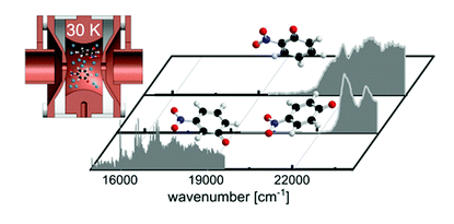
The intrinsic photophysics of nitrophenolate isomers (meta, para, and ortho) was studied at low temperature using photodissociation mass spectrometry in a cryogenic ion trap instrument. Each isomer has distinct photophysics that affects the excited state lifetimes, as observed experimentally in their spectroscopic linewidths. Visible-light-induced excitation of m-nitrophenolate gives rise to well-resolved vibronic features in the spectrum of the S1 state. The para and ortho isomers have broad spectra – even at cryogenic temperatures – due to their shorter excited state lifetimes and spectral congestion. We present computational evidence for mixing of the first and second excited states of o-nitrophenolate, leading to significant additional broadening in the experimental spectrum.

DOI: 10.1039/C8CP06078A

Leah G. Dodson,a  Wyatt Zagorec-Marks,b  Shuang Xu,c  James E. T. Smithb  and  J. Mathias Weberb  

aJILA and NIST, University of Colorado, 0440 UCB, Boulder, USA
bJILA and Department of Chemistry, University of Colorado, 0440 UCB, Boulder, USA
cJILA and Department of Physics, University of Colorado, 0440 UCB, Boulder, USA
Electronic Spectra of Tris (2, 2′-bipyridine)-M (II) Complex Ions in vacuo (M= Fe and Os)

We measured the electronic spectra of mass-selected [M(bpy)3]2+ (M = Fe and Os, bpy = 2,2′-bipyridine) ions in vacuo by photodissociation spectroscopy of their N2 adducts, [M(bpy)3]2+·N2. Extensive band systems in the visible (predominantly charge transfer) and near-ultraviolet (ππ*) spectral regions are reported. The [M(bpy)3]2+·N2 target ions were prepared by condensing N2 onto electrosprayed ions in a cryogenic ion trap at ca. 25 K and then mass-selected by time-of-flight mass spectrometry. The electronic photodissociation spectra of the cold, gas-phase ions closely reflect their intrinsic properties, i.e., without perturbation by solvent effects. The spectra are interpreted using time-dependent density functional theory calculations both with and without accounting for relativistic effects.

DOI: 10.1021/acs.inorgchem.7b00620

Shuang Xu, James E. T. Smith, and J. Mathias Weber

JILA and Department of Physics, University of Colorado, Boulder, Colorado 80309-0440, United States
JILA and Department of Chemistry and Biochemistry, University of Colorado, Boulder, Colorado 80309-0440, United States

UV Spectra of Tris (2, 2′-bipyridine)–M (II) Complex Ions in vacuo (M= Mn, Fe, Co, Ni, Cu, Zn)

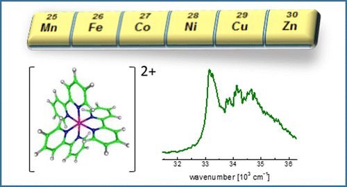
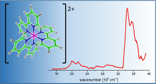
We present electronic spectra in the π–π* region of a series of tris(bpy)–M(II) complex ions (bpy = 2,2′-bipyridine; M = Mn, Fe, Co, Ni, Cu, Zn) in vacuo for the first time. By applying photodissociation spectroscopy to cryogenically cooled and mass selected [MII(bpy)3]2+ ions, we obtain the intrinsic spectra of these ions at low temperature without perturbation by solvent interaction or crystal lattice shifts. This allows spectroscopic analysis of these complex ions in greater detail than possible in the condensed phase. We interpret our experimental data by comparison with time-dependent density functional theory.

DOI: 10.1021/acs.inorgchem.6b02054

Shuang Xu, James E. T. Smith, and J. Mathias Weber

JILA and Department of Physics, University of Colorado, Boulder, Colorado 80309-0440, United States
JILA and Department of Chemistry and Biochemistry, University of Colorado, Boulder, Colorado 80309-0440, United States

Hydration of a Binding Site with Restricted Solvent Access: Solvatochromic Shift of the Electronic Spectrum of a Ruthenium Polypyridine Complex, One Molecule at a Time

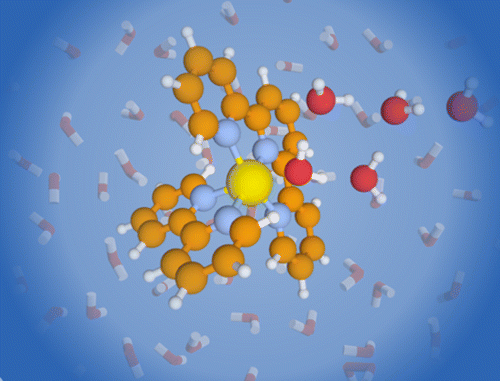
We report the electronic spectra of mass selected [(bpy)(tpy)Ru–OH2]2+·(H2O)n clusters (bpy = 2,2′-bipyridine, tpy =2,2′:6′2″-terpyridine, n = 0–4) in the spectral region of their metal-to-ligand charge transfer bands. The spectra of the mono- and dihydrate clusters exhibit partially resolved individual electronic transitions. The water network forming at the aqua ligand leads to a rapid solvatochromic shift of the peak of the band envelope: addition of only four solvent water molecules can recover 78% of the solvatochromic shift in bulk solution. The sequential shift of the band shows a clear change in behavior with the closing of the first hydration shell. We compare our experimental data to density function theory (DFT) calculations for the ground and excited states.

DOI:  10.1021/acs.jpca.6b07668

Shuang Xu, James E. T. Smith, and J. Mathias Weber

 JILA and Department of Physics, University of Colorado, Boulder, Colorado 80309-0440, United States
 JILA and Department of Chemistry and Biochemistry, University of Colorado, Boulder, Colorado 80309-0440, United States

The electronic spectrum of cryogenic ruthenium-tris-bipyridine dications in vacuo

We report the electronic spectrum of the prototypical ruthenium coordination complex Ru(bpy) 2+ (bpy = 2, 2′-bipyridine) by messenger tagging with N in a cryogenic ion trap and photodissociation spectroscopy of mass selected Ru(bpy) 2+ ⋅ N ions. We observe individual electronic bands and groups of bands with unprecedented detail, particularly in the usually unresolved metal-to-ligand charge transfer region of thespectrum. By comparing our experimental results with time-dependent density functional theory, both with and without spin-orbit interaction [Heully , J. Chem. Phys. , 184308 (2009)], we are able to assign the spectrum of the isolated ion.

DOI:  10.1063/1.4955262

Shuang Xu, James E. T. Smith, and J. Mathias Weber

JILA and Department of Physics, University of Colorado, Boulder, Colorado 80309-0440, United States
JILA and Department of Chemistry and Biochemistry, University of Colorado, Boulder, Colorado 80309-0440, United States

Ligand Influence on the Electronic Spectra of Dicationic Ruthenium-Bipyridine-Terpyridine Complexes

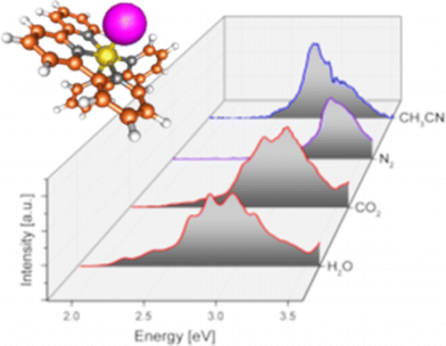
We report electronic spectra of a series of ruthenium polypyridine complexes of the form [(trpy)(bipy)RuII–L]2+ (bipy = 2,2′-bipyridine and trpy = 2,2′:6′,2″-terpyridine), where L represents a small molecular ligand that occupies the last coordination site. Species with L = H2O, CO2, CH3CN, and N2 were investigated in vacuo using photodissociation spectroscopy. All species exhibit bright metal-to-ligand charger transfer (MLCT) bands in the visible and near UV, but with different spectral envelopes and peak energies, encoding the influence of the ligand L on the electronic structure of the complex. Several individual electronic bands can be resolved for L = H2O and CO2, while the spectra for L = N2 and CH3CN are more congested, even at low ion temperatures. The experimental results are discussed in the framework of time-dependent density functional theory.

DOI: 10.1021/acs.jpca.6b02926

Shuang Xu, James E. T. Smith, and J. Mathias Weber

JILA and Department of Physics, University of Colorado, Boulder, Colorado 80309-0440, United States
JILA and Department of Chemistry and Biochemistry, University of Colorado, Boulder, Colorado 80309-0440, United States

Absorption Spectrum of a Ru(II)-Aquo Complex in Vacuo: Resolving Individual Charge-Transfer Transitions

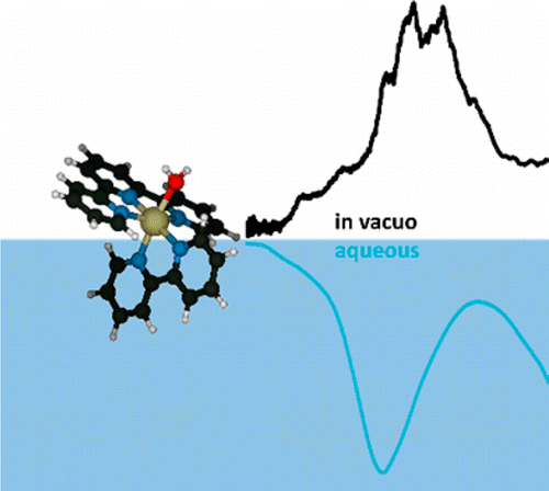
Ruthenium(II) complexes are of great interest as homogeneous catalysts and as photosensitizers; however, their absorption spectra are typically very broad and offer only little insight into their electronic structure. We present the electronic spectrum of the aquo complex [(trpy)(bipy)RuII–OH2]2+ measured by photodissociation spectroscopy of mass-selected ions in vacuo (bipy = 2,2′-bipyridine and trpy = 2,2′:6′,2″-terpyridine). In the visible and near-UV, [(trpy)(bipy)RuII–OH2]2+ has several electronic bands that are not resolved in absorption spectra of this complex in solution but are partially resolved in vacuo. The experimental results are compared with results from time-dependent density functional theory calculations.
Shuang Xu and J. Mathias Weber*§
JILA, University of Colorado, 440 UCB, Boulder, Colorado 80309, United States
Department of Physics, University of Colorado, Boulder, Colorado 80309, United States
§ Department of Chemistry and Biochemistry, University of Colorado, Boulder, Colorado 80309, United States
 

Ligand influence on the electronic spectra of monocationic copper–bipyridine complexes

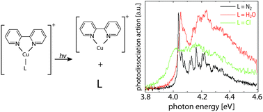
We present photodissociation spectroscopy and computational analysis of three monocationic Cu–bipyridine complexes with one additional ligand of different interaction strength (N2, H2O and Cl) in the visible and UV. All three complexes show similar ππ* bands with origins slightly above 4 eV and vibrational band contours that are due to bipyridine ring deformation modes. Experiments at low temperature show that excited-state lifetime is the limiting factor for the width of the vibrational features. In the case of Cl as a ligand, there is a lower lying bright ligand-to-ligand charge-transfer state around 2.75 eV. The assignment of the transitions was made based on equation-of-motion coupled-cluster calculations. While the nature of the ligand does not significantly change the position of the bright ππ* state, it drastically changes the excited-state dynamics.
 

DOI: 10.1039/C5CP05063D

Shuang Xu,a   Samer Gozem,b   Anna I. Krylov,b  Casey R. Christopherc and   J. Mathias Weber*c
 aJILA and Department of Physics, University of Colorado, Boulder, USA
bDepartment of Chemistry, University of Southern California, Los Angeles, USA
cJILA and Department of Chemistry and Biochemistry, University of Colorado, Boulder, USA
* weberjm@jila.colorado.edu

Landau-Zener-Stückelberg spectroscopy of a superconducting flux qubit

We proposed a different method to measure the energy spectrum of a superconducting flux qubit. Different from the conventional frequency spectroscopy, a linear triangle pulse was used to drive the qubit through the anticrossing and to generate Landau-Zener-Stückelberg interference patterns, from which the information of the energy spectrum can be extracted. This method facilitates the spectroscopy in regimes of very large energies. Moreover, without installing microwaves lines one can simplify the experimental setup and reduce the unwanted effects of noise. The idea can be applied to other quantum systems as well, opening the possibility of calibrating and manipulating qubits with linear pulses.

DOI: PhysRevB.82.144526

Shuang Xu and Yang Yu*
National Laboratory of Solid State Microstructures and Department of Physics, Nanjing University, Nanjing 210093, China
Guozhu Sun
Institute of Superconductor Electronics and Department of Electronic Science and Engineering, Nanjing University, Nanjing 210093, China
*yuyang@nju.edu.cn